
The relentless itching isn’t a sign of a flea infestation; it’s an extreme allergic reaction to proteins in a single flea’s saliva.
- For a hypersensitive dog, the problem is not the flea itself, but the immunological crisis it triggers, which can last for weeks.
- Effective control means killing fleas before they can bite and simultaneously treating the dog’s overactive immune response.
Recommendation: Shift your focus from “finding fleas” to implementing a veterinary-guided plan that combines rapid-kill preventatives with anti-itch therapies to break the cycle.
As a veterinary dermatologist, the most common and frustrating statement I hear from owners of intensely itchy dogs is, “But Doctor, it can’t be fleas. I’ve never seen a single one.” This is the central paradox of Flea Allergy Dermatitis (FAD), a condition where the visible evidence (or lack thereof) is profoundly misleading. The problem isn’t a house crawling with parasites; it’s a severe, localized immunological crisis triggered by an invisible encounter. One flea, in one brief moment, can bite, feed, and leave, injecting a cocktail of antigenic proteins into your dog’s skin.
For a normal dog, this is a minor annoyance. For a dog with FAD, it’s a declaration of war. Their immune system doesn’t just react; it overreacts violently. This hypersensitivity is what transforms a fleeting bite into weeks of chewing, scratching, hair loss, and misery. The flea is long gone, but the inflammatory cascade it initiated rages on. This is why you don’t see the culprit, only the collateral damage. Your dog isn’t suffering from a flea problem; they are suffering from an allergy, and it’s a medical condition that requires a specific, multi-faceted strategy.
Understanding this distinction is the first step toward true relief. It’s not about flea bombs and powders alone. It’s about interrupting the body’s allergic response, managing the secondary skin damage, and deploying a preventative so fast it neutralizes the threat before the allergic trigger can even be pulled. This guide will walk you through the clinical signs, the medical tools to stop the itch, and the strategic prevention necessary to manage this chronic condition effectively.
To navigate this complex issue, we will break down the key components, from identifying the tell-tale signs of the allergy to understanding the nuances of treatment and prevention. This framework will provide the clarity needed to finally bring your dog relief.
Summary: A Dermatologist’s Guide to Flea Allergy Dermatitis
- The “Tail Base” Pattern: Recognizing the Classic Flea Allergy Sign
- Steroids vs. Apoquel: How to Stop the Scratching Cycle Quickly?
- Anxiety and Aggression: How Chronic Itch Changes Behavior?
- Oral Tablets vs. Topicals: Which Kills Before the Flea Bites?
- Hot Spots: How to Treat the Wet Scabs Caused by Scratching?
- The Pupa Stage: Why Fleas Return 2 Weeks After Treatment?
- Scissors vs. Clippers: Why Cutting Mats Is Dangerous?
- Coffee Grounds in Ears: Is It Dirt or Ear Mites?
The “Tail Base” Pattern: Recognizing the Classic Flea Allergy Sign
While you may not see the flea, you can absolutely see the evidence it leaves behind on a hypersensitive dog. Flea Allergy Dermatitis isn’t a random, all-over itch. It presents in a classic, tell-tale distribution that veterinarians call the “flea triangle” or “pants pattern.” The reaction is most concentrated on the lower back, the base of the tail, and down the back of the hind legs. This area becomes the epicenter of your dog’s misery, leading to frantic chewing, hair loss, and inflamed skin.
This distinct pattern is a powerful diagnostic clue. The reason for this specific location is thought to be related to flea behavior; they often congregate around the tail and rump. In dogs, this manifests as lesions primarily on the lower back, tailhead, and inner thighs. The skin may be red, bumpy, and thickened from chronic scratching, a condition known as lichenification. You may also see small scabs (papules) and significant hair loss. In fact, Flea allergy dermatitis is the most common dermatologic disease of domestic dogs in the US, making recognition of this pattern crucial.
This visual below shows the typical area a veterinarian examines to confirm suspicions of FAD. The focused inflammation is a hallmark of the condition.
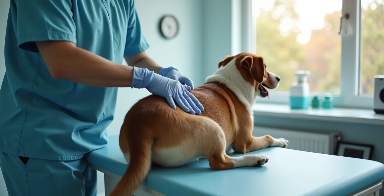
Seeing this pattern, even in the complete absence of visible fleas, is a strong indicator that FAD is the underlying problem. The self-trauma is a direct result of the intense, localized pruritus (itch) caused by the allergic reaction to flea saliva. Recognizing this specific distribution is the first step in getting the correct diagnosis and breaking the cycle of misery for your pet.
Steroids vs. Apoquel: How to Stop the Scratching Cycle Quickly?
When a dog is in the throes of an FAD flare-up, the primary goal is to stop the itch-scratch cycle as quickly as possible. This is not just for comfort; it’s to prevent the self-trauma that leads to secondary infections and chronic skin changes. The two mainstays for rapid itch relief are corticosteroids (like prednisone) and targeted therapies like oclacitinib (Apoquel). As a dermatologist, my choice depends on the severity of the flare-up and the long-term plan.
Corticosteroids are powerful, broad-spectrum anti-inflammatories. They work by suppressing the entire immune system, which effectively shuts down the allergic reaction. They are fast-acting, often providing relief within hours, making them excellent for severe, acute flare-ups. However, their broad action comes with a higher risk of side effects, including increased thirst and urination, and they are not ideal for long-term use. Abruptly stopping steroids can also lead to a severe rebound of itching.
Apoquel (oclacitinib), on the other hand, is not a steroid. It is a Janus kinase (JAK) inhibitor that specifically targets the signaling pathways that cause itch and inflammation. It’s more like a guided missile than a bomb. It provides rapid relief, often within 4 hours, but with a more targeted action and generally fewer side effects than steroids, making it a preferred choice for long-term management. The following table from an analysis of allergy medications compares these options.
| Medication | Mechanism | Onset | Duration | Best For |
|---|---|---|---|---|
| Steroids (Prednisone) | Suppresses entire immune system | Hours | Short-term only | Severe acute flare-ups |
| Apoquel (Oclacitinib) | Targets JAK/STAT pathways | 4 hours | Daily dosing | Long-term management |
| Cytopoint (Lokivetmab) | Neutralizes IL-31 protein | 1-2 days | 4-8 weeks | Compliance issues, long-term relief |
Often, a clinical strategy involves using a short, tapering course of steroids to get a severe flare-up under control, while simultaneously starting a long-term medication like Apoquel or Cytopoint. This provides immediate relief and creates a bridge to a safer, sustainable management plan.
Action plan: Managing the rebound effect when stopping steroids
- Never stop steroids abruptly – always follow veterinary tapering schedule.
- Begin transitioning to Apoquel or Cytopoint before completing the steroid taper.
- Maintain strict flea prevention throughout the transition period.
- Monitor for increased scratching during dose reductions.
- Contact your veterinarian if severe itching returns before the next scheduled dose.
Anxiety and Aggression: How Chronic Itch Changes Behavior?
One of the most overlooked consequences of FAD is its profound impact on a dog’s behavior and mental state. The relentless, inescapable itch is more than a simple annoyance; it’s a form of chronic pain and stress. This constant state of discomfort can fundamentally alter your dog’s personality, leading to behaviors that owners may mistakenly attribute to poor training or temperament.
Dogs suffering from chronic pruritus often experience poor sleep quality, leading to irritability and a decreased threshold for frustration. They may become restless, unable to settle, and constantly preoccupied with chewing or scratching. This can manifest as anxiety, obsessive-compulsive behaviors, or even aggression. A dog that was once placid and friendly may begin to snap when touched near sensitive areas or become generally more reactive to other pets and people in the household.
Chronic pruritus is not just an annoyance but a form of pain that elevates cortisol (the stress hormone), leading to poor sleep, irritability, and a lower threshold for aggression.
– Veterinary Dermatology Specialists, Haarstad Veterinary Dermatology
This behavioral decline can severely strain the human-animal bond. A study of dogs with chronic FAD highlighted that the pet’s constant discomfort and resulting behavioral issues cause significant stress for owners. Many reported frustration and a feeling of helplessness, with some even considering behavioral euthanasia before the underlying FAD was properly diagnosed and treated. Addressing the itch is not just about healing the skin; it is about restoring the dog’s quality of life and preserving the precious relationship with its family. When the itch is controlled, owners are often amazed to see their “old dog” come back—calmer, happier, and more engaged.
Oral Tablets vs. Topicals: Which Kills Before the Flea Bites?
For a non-allergic dog, any effective flea product will eventually solve the problem. For a dog with FAD, “eventually” is not good enough. The entire strategy hinges on killing the flea before it has a chance to bite and inject its allergenic saliva. This is where the speed of kill becomes the single most important factor in choosing a preventative. A product that takes 24 hours to kill a flea is 23 hours too slow for a hypersensitive patient.
This is why modern oral medications, specifically those in the isoxazoline class (e.g., Bravecto, NexGard, Simparica, Credelio), have become the gold standard for managing FAD. Unlike many older topical products that can take hours or days to achieve full efficacy, these oral tablets are absorbed into the bloodstream and work incredibly fast. According to veterinary dermatology specialists, isoxazoline tablets can start killing fleas as quickly as 30 minutes to 8 hours after ingestion. This rapid action means the flea dies shortly after its first bite, often before it can inject enough saliva to trigger a full-blown allergic reaction.
Topical (spot-on) treatments can still be effective, but they have more variables that can lead to failure. For a topical to work, it must be applied correctly to the skin, not just the hair, and the dog should not be bathed for at least 48 hours after application. Furthermore, some flea populations have developed resistance to older topical ingredients like fipronil. If you’re using a topical and still seeing flare-ups, it’s crucial to troubleshoot the application or discuss switching to an oral product with your veterinarian. For FAD management, the goal is not just to kill fleas, but to create a “no-bite zone” around your dog, and fast-acting oral tablets are the most reliable way to achieve this.
Hot Spots: How to Treat the Wet Scabs Caused by Scratching?
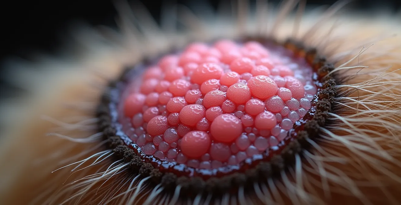
“Hot spots,” known in veterinary terms as acute moist dermatitis or pyotraumatic dermatitis, are a frequent and painful complication of FAD. They are not a separate disease, but a direct result of the intense, focused self-trauma caused by the itch. A dog will chew or scratch a single spot so aggressively that it erodes the surface of the skin, creating a weeping, raw, and infected lesion that can appear and expand dramatically within hours.
The initial skin damage allows bacteria and yeast that normally live on the skin to overgrow, leading to a secondary infection. In cases of severe FAD, these lesions are often complicated by Staphylococcus bacterial infections and Malassezia yeast overgrowth. These microbes can create a protective biofilm that resists simple cleaning, making the infection harder to treat. This is why a hot spot is more than just a scab; it’s an active, painful infection that requires prompt medical intervention to resolve. The goal of treatment is twofold: stop the itching that caused it and treat the secondary infection.
This close-up image shows the texture of a healing hot spot, where new, healthy pink tissue is forming at the center while the edges remain crusted.
While veterinary care is essential for proper treatment, you can perform first aid at home to prevent a small spot from worsening. The protocol involves three key steps:
- Clip the hair: Carefully clip the fur around the lesion to allow air to reach the skin. This prevents moisture from being trapped and helps medication make direct contact.
- Clean the area: Gently clean the surface with a veterinary-approved antiseptic solution, such as one containing chlorhexidine. Do not use soap, hydrogen peroxide, or alcohol, as these can damage healing tissue.
- Prevent further licking: This is the most critical step. Use an Elizabethan collar (e-collar) to stop your dog from licking or chewing the area, which would prevent healing and worsen the infection. Your veterinarian will then prescribe appropriate topical and sometimes oral antibiotics or antifungals.
The Pupa Stage: Why Fleas Return 2 Weeks After Treatment?
One of the biggest frustrations for owners is the seeming recurrence of fleas weeks after starting a treatment. You’ve treated your dog, the itching has improved, and then suddenly, the problem is back. This isn’t a treatment failure; it’s the result of the flea life cycle unfolding in your home. The adult fleas you see on your pet represent only 5% of the total flea population. The other 95% exist as eggs, larvae, and pupae hidden in your carpets, bedding, and furniture.
The pupal stage is the most formidable part of this cycle. After the larva spins its cocoon, it becomes a pupa. This sticky, resilient casing is virtually indestructible. It is resistant to insecticides, freezing temperatures, and drying. The pre-emerged adult flea can remain dormant inside this protective shell for weeks or even months, waiting for the right signal to emerge: the vibration, heat, and carbon dioxide from a passing host—your dog.
This is why you can have a “flea gap.” You kill all the adult fleas on your pet, but two weeks later, a new generation of fleas emerges from the pupae in the environment and re-infests your dog. As UC Davis Veterinary Medicine reports, for one adult flea found on your pet there are at least one hundred immature fleas in the animal’s environment. Breaking the cycle requires persistence.
Eliminating a household flea population is not a one-time event; it’s a process that takes at least three months. The following timeline illustrates what is happening behind the scenes.
| Month | What’s Happening | What You’ll See | Action Needed |
|---|---|---|---|
| Month 1 | Adult fleas killed, pupae still hatching | May still see some fleas | Continue treatment, vacuum frequently |
| Month 2 | Fewer pupae hatching, eggs prevented | Significant reduction in fleas | Maintain prevention, treat environment |
| Month 3 | Cycle broken | No visible fleas | Continue year-round prevention |
This is why year-round, uninterrupted flea prevention is non-negotiable for an allergic dog. You must keep a fast-acting preventative on board continuously to kill these newly hatched fleas before they have a chance to bite.
Scissors vs. Clippers: Why Cutting Mats Is Dangerous?
When a dog’s skin is inflamed and itchy from FAD, the condition of their coat becomes critically important. The constant scratching and chewing can cause the fur to become tangled and matted, especially in longer-haired breeds. These mats are not just an aesthetic problem; they are a significant medical risk that can dramatically worsen skin disease.
Mats trap moisture, heat, and dirt against already inflamed skin, creating a perfect breeding ground for the secondary infections that cause hot spots.
– Professional Grooming Association, Veterinary Dermatology Guidelines
A common but dangerous impulse for owners is to try and cut these mats out with scissors. This is extremely hazardous. Mats often form very tightly against the skin, and because the skin underneath is often inflamed and swollen, it can easily be pulled up into the mat. It is incredibly common for owners (and even inexperienced groomers) to accidentally cut the skin while trying to cut out a mat, leading to a serious laceration that requires stitches.
The safest way to remove mats is with electric clippers using a short blade (like a #10 blade). The clippers are designed to glide along the skin’s surface, getting underneath the mat and shaving it off without the risk of cutting the skin itself. For small knots, you may be able to work them out with your fingers and a detangling spray. For medium mats, a specialized tool called a mat-splitter can be used with care. However, for severe or widespread matting, the only safe options are electric clippers, either at a professional groomer or your veterinary clinic. Never risk using scissors on a mat—the danger of causing a severe injury is far too high.
Key takeaways
- Flea Allergy Dermatitis is an immunological overreaction to flea saliva, not a simple infestation.
- The classic sign is intense itching and hair loss on the lower back, tail base, and hind legs.
- Effective management requires both rapid-kill flea prevention (ideally oral) and veterinary medications to control the allergic reaction.
Coffee Grounds in Ears: Is It Dirt or Ear Mites?
When a dog has FAD, the inflammation is not always confined to the “flea triangle.” The systemic allergic reaction can compromise the skin barrier all over the body, including inside the ear canals. This makes the dog more susceptible to secondary ear infections, a problem often mistaken by owners for ear mites. A common sign is a dark, granular discharge that resembles coffee grounds.
While ear mites (Otodectes cynotis) do produce this characteristic dark, crumbly debris, it is a far less common diagnosis in adult dogs than a secondary infection with yeast or bacteria. A yeast infection, typically from Malassezia, is extremely common in allergic dogs and produces a dark brown, greasy or waxy paste with a distinct sweet, musty odor. Bacterial ear infections can also produce a dark discharge, though it is often more yellowish and liquid, with a foul smell. As a case study on FAD notes, the widespread inflammation compromises the ear canal’s skin barrier, predisposing pets to these secondary infections that require different treatments than ear mites.
Attempting to treat a yeast or bacterial infection with an over-the-counter ear mite medication will be completely ineffective and will only delay proper treatment, allowing the infection and pain to worsen. A definitive diagnosis can only be made by your veterinarian, who will take a sample of the debris and examine it under a microscope. This simple test, called cytology, quickly reveals whether the culprit is mites, yeast, bacteria, or a combination.
The table below, based on clinical findings, outlines the key differences a veterinarian looks for to distinguish between these common causes of dark ear debris.
| Condition | Appearance | Consistency | Odor | Microscopic Finding |
|---|---|---|---|---|
| Ear Mites | Dark brown ‘coffee grounds’ | Dry, crumbly | Minimal | Visible mites under oil |
| Yeast Infection | Dark brown paste | Greasy, waxy | Sweet, musty | Malassezia organisms |
| Bacterial Infection | Yellowish/pus-like | Liquid to thick | Foul | Cocci or rods on stain |
Ultimately, managing Flea Allergy Dermatitis requires a paradigm shift. It’s about moving away from the hunt for a visible pest and toward the clinical management of a chronic allergic disease. By partnering with your veterinarian, you can create a comprehensive plan that stops the itch at its source, treats the secondary damage, and deploys an impenetrable defense against future bites. This proactive, medical approach is the only way to give your hypersensitive dog the lasting peace and comfort they deserve.